Early pregnancy scan

An early pregnancy scan is usually the first ultrasound examination done once the pregnancy is suspected (missed period) and confirmed by a urine pregnancy test (UPT).This scan confirms the presence of pregnancy and its location whether it is inside or outside the uterus.

The number of babies,the chorionicity in case of multiple gestation and the age of pregnancy is determined followed by confirmation of its viability.

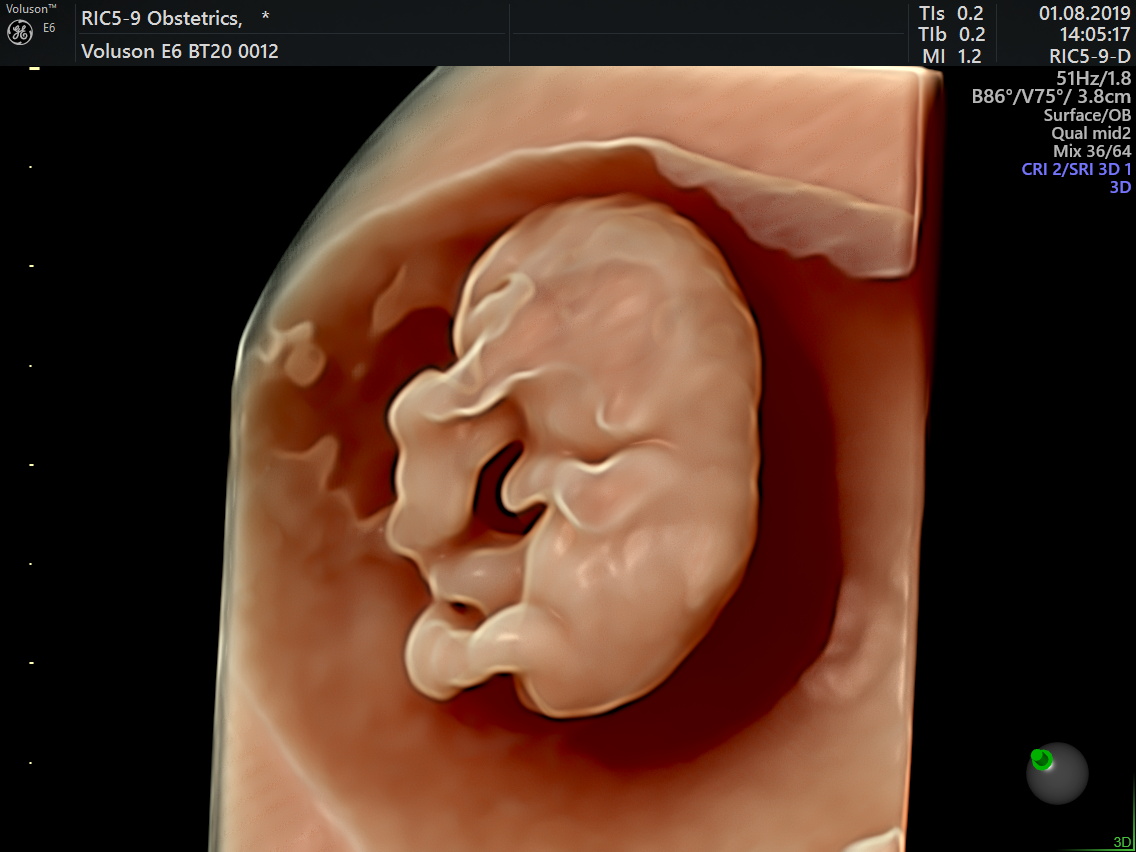
First Trimester Screening

An effective method of screening is based in the combination of:

Maternal age

A maternal blood sample for the measurement of free ß-hCG and PAPP-A(Double marker test)

An ultrasound scan (NT scan) at 11-13 weeks +6 days (preferably after 12 weeks).

To measure the collection of fluid behind the fetal neck (nuchal translucency)

To examine the fetal nasal bone and palate

To measure the fetal heart rate

To assess the flow of blood across the tricuspid valve of the fetal heart and the ductus venosus

This method of screening increases the detection rate of Down’s syndrome and other major chromosomal abnormalities from less than 50% to more than 95%.

Other benefits of the 11–13 weeks scan include:

Accurate dating of the pregnancy

Early diagnosis of many major fetal abnormalities

The detection of multiple pregnancies with reliable diagnosis of chorionicity, which is the main determinant of the outcome in multiple pregnancies

Another recent development is that the 11-13 weeks scan can be used to identify women at increased risk for the development of preeclampsia in pregnancy

Screening for Preeclampsia

It includes assessment of maternal mean arterial blood pressure and uterine artery Doppler.

The objective of screening for PE at 11-13 weeks’ gestation is to identify the cases that would benefit from prophylactic use of aspirin that reduces the risk of preterm PE by more than 60%

The objective of screening for PE at 20-24 weeks’ gestation is to estimate the patient-specific risk of developing PE and on the basis of such risk define the subsequent management of pregnancy, including the timing and content of subsequent visits. This would potentially minimize adverse perinatal events for those that develop PE by determining the appropriate time and place for delivery.

Mid-trimester Anomaly Scan/Targeted Imaging for Fetal Anomalies (TIFFA scan)

As the name suggests this is a targeted scan performed ideally at 18-24 weeks. The aim is to study all the structures and organs of the baby and assess the baby for any structural abnormality.

The study also focuses on screening for chromosomal abnormalities by looking for “Soft Markers“. A basic yet an in-depth examination of fetal heart is an integral part of this scan. Reassessment of risk of pre-eclampsia and FGR is also performed during this scan by assessing the uterine artery Doppler parameters.

An early anomaly scan between 16-18 weeks may be indicated in some cases where an abnormality is suspected in the first trimester or in cases with previous history of fetal abnormalities.

Growth Scan

Performed from 26-28 weeks onwards, the study focuses on detailed assessment of the growth and well being of the fetus in order to diagnose babies who are unable to attain their actual growth potential.

This scan is also important to diagnose evolving abnormalities,especially heart and brain anomalies.

Doppler Scan

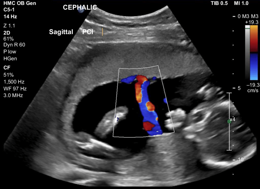
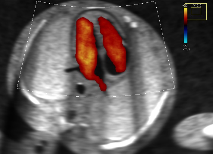
This includes assessment of maternal and fetal blood flows in case of suspected growth restriction. Timely intervention and management can then be offered in case of fetal growth restriction, abnormal blood flow and reduced amniotic fluid.

Fetal and maternal Doppler assessment is an integral part of a growth scan .

Fetal Echocardiography

It is a detailed structural and functional study of the baby’s heart (apart from the cardiac examination performed at the time of anomaly scan) performed between 18-24 weeks of pregnancy. Ideally recommended in mothers with high risk of baby with heart disease or suspicion of fetal heart disease in previous scan.

Fetal Neurosongraphy

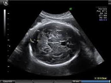
Central nervous system (CNS) malformations are some of the most common of all congenital abnormalities.Neurosonography includes multiplanar imaging of fetal brain in fetuses at risk for acquired brain anomalies

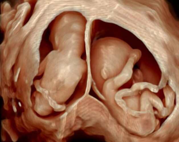
Multiple Pregnancy Scan and Evaluation

Multiple pregnancy is a condition in which more than one fetus develops simultaneously in the mother’s womb.Multifetal pregnancies account for ~1% of all pregnancies but are seen in much higher numbers in populations where in vitro fertilisation (IVF) is a common practice.

All twin pregnancies have higher fetal and neonatal mortality with overall rates of fetal mortality being 3-6 times that of a singleton pregnancy and neonatal mortality 7 times that of single pregnancies.

Monochorionic twin pregnancies share the one placenta and are therefore prone to hemodynamic complications such as twin-twin transfusion (TTTS),twin anemia polycythemia sequence (TAPS),twin reversed arterial perfusion sequence (TRAPS),acardiac twin,demise of co twin. The role of ultrasound is crucial in the monitoring of the pregnancy and planning for delivery.

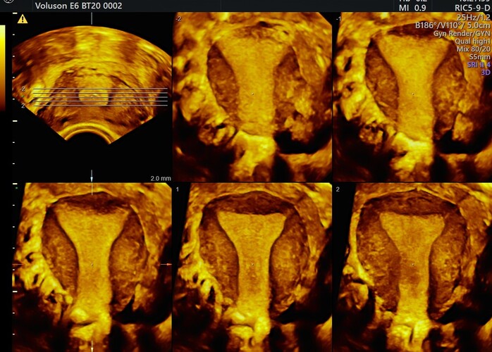
Gynaecological Scans (3D scan)

Gynaecological scans are used to assess organs and structures within the female pelvis.

A pelvic ultrasound allows quick visualization of the female pelvic organs and structures including the uterus, cervix, vagina, fallopian tubes and ovaries.

A pelvic ultrasound may be used to diagnose and assist in the treatment of the following conditions:

Abnormalities in the anatomic structure of the uterus(uterine anomalies-3D) including endometrial conditions.

Fibroid tumors (benign growths), masses, cysts, and other types of tumors within the pelvis.

Ovarian/Adnexal masses.

Presence and position of an intrauterine contraceptive device (IUD)

Pelvic inflammatory disease (PID) and other types of inflammation or infection

Monitoring of ovarian follicle size for infertility evaluation

Ectopic pregnancy (pregnancy occurring outside of the uterus, usually in the fallopian tube)

Assessing certain fetal conditions

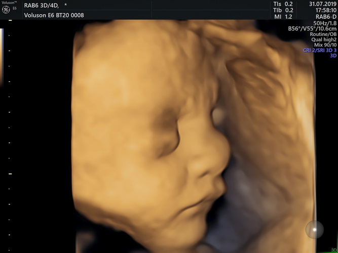
3D/4D Scans

3D scans show still pictures of your baby in three dimensions. 4D scans show moving 3D images of your baby. The best time to obtain good 3D/4D images of your baby is between 24 and 37 weeks.